Tooth Attrition - What It Is, What Causes It, and How to Treat It
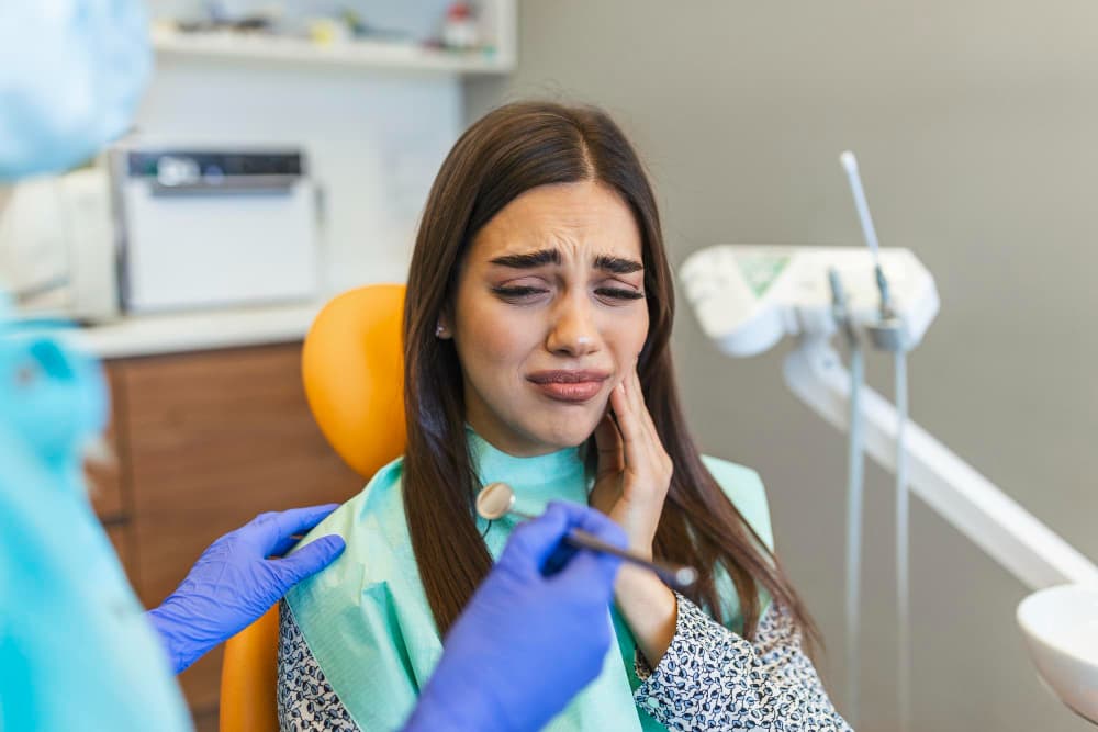
Young woman holding cheek in chair at dentist having toothache shot.
Tooth wear is not a single condition — it's a family of conditions, each driven by different mechanisms. Attrition is the specific type of tooth wear caused by direct tooth-to-tooth contact. Unlike erosion (chemical wear from acid) or abrasion (wear from external objects like hard toothbrushes), attrition happens from the inside — from the mechanical grinding of the upper and lower teeth against each other.
The result is predictable: the biting edges and chewing surfaces of the teeth become progressively flatter and shorter. In mild cases, the change is cosmetic. In severe cases, it alters the entire vertical dimension of the bite, affects jaw joint function, and requires extensive and costly rehabilitation.
At Renew Dental Clinic in Sector 47, Noida, tooth wear — including attrition — is one of the conditions Dr. Suchi Singh assesses at every check-up, because the consequences of untreated progressive attrition worsen over time in a way that early management could prevent.
What Is Dental Attrition?
The word "attrition" comes from the Latin for "wearing away by friction." In dentistry, it refers to the loss of tooth structure that occurs specifically from contact between the opposing (upper and lower) teeth during chewing, swallowing, or grinding.
Every tooth in the mouth has an opposing tooth — a partner it meets when you bite or chew. Under normal eating loads, this contact causes minimal wear over a lifetime because the duration and force of contact are physiologically controlled. The problem arises when contact is excessive — either because of parafunctional habits like bruxism (grinding or clenching) or because of a bite that places abnormal loading on specific teeth.
Attrition is characterised by:
- Flat, polished surfaces on the biting edges and cusp tips — the characteristic "worn" appearance of teeth that have lost their normal anatomy
- Matching wear facets on opposing teeth — the worn surfaces on an upper tooth correspond precisely to the worn surface on the lower tooth it contacts
- Progressive shortening of the teeth over time
Causes of Dental Attrition
Bruxism — The Primary Cause
Bruxism — the involuntary grinding or clenching of teeth — is the leading cause of pathological attrition. During sleep bruxism, the jaw muscles generate forces that are many times greater than those of normal chewing, applied in lateral (side-to-side) movements rather than the vertical force of a normal bite.
These lateral forces are particularly damaging because they cause teeth to slide against each other over their entire contact surface rather than merely tapping together briefly. The cumulative effect over months and years is significant, visible enamel loss from all surfaces that come into contact.
Sleep bruxism is often undiagnosed because it occurs during sleep — the patient has no memory of it. Signs at check-up include characteristic flat facets, enamel thinning, and often jaw muscle tenderness on palpation.
Normal Ageing and Physiological Wear
A degree of tooth wear is entirely normal over a lifetime. Decades of chewing will produce some reduction in the height of the biting surfaces of the back teeth — this is physiological attrition. Its rate depends on diet hardness, chewing habits, and the natural hardness of the patient's enamel.
Physiological attrition is slow, gradual, and generally doesn't require treatment beyond monitoring. The concern is when wear rate is accelerated beyond what is expected for the patient's age — which suggests a pathological factor like bruxism is involved.
Malocclusion (Bite Misalignment)
When the teeth don't meet in their ideal alignment, certain teeth bear disproportionate contact. These teeth experience accelerated attrition because they're absorbing contact forces that should be shared across more surfaces. Crossbite, deep overbite, and other bite problems concentrate loading in ways that accelerate wear on specific teeth.
Diet — Hard or Coarse Foods
Populations that traditionally eat coarsely ground grains, hard foods, or gritty foods show higher rates of generalised attrition — because the food particles trapped between teeth during chewing act as a mild abrasive on the contact surfaces. This is less significant in modern urban diets where food processing removes most coarse particles, but remains relevant for patients whose diet includes a high proportion of hard, tough foods eaten regularly.
Acid Combined with Attrition (Combined Wear)
Enamel softened by acid exposure — from dietary acid, GERD, or frequent vomiting — is significantly more susceptible to mechanical wear. In patients who have both acid exposure and bruxism, the combined rate of tooth wear is dramatically higher than either factor alone would produce. This combined wear pattern is increasingly common and requires management of both the acid exposure and the mechanical force simultaneously.
What Attrition Does to the Teeth — Progressive Consequences
Early Stage — Enamel Loss
The first wear occurs in the enamel layer. As enamel is flattened, the natural cusp anatomy disappears — the rounded, raised cusps of the back teeth become flat, and the incisal edges of the front teeth become straight rather than naturally curved.
At this stage, the teeth may begin to feel slightly different but often cause no discomfort. The change may only be noticed at a dental check-up.
Dentine Exposure — Sensitivity Develops
Once enamel wear reaches the dentine layer, sensitivity typically begins. Dentine contains microscopic tubules connected to the nerve — exposure of dentine produces sensitivity to cold, air, and sometimes sweet foods. This is a clear signal that wear has progressed beyond the enamel.
The exposed dentine also wears significantly faster than enamel — the rate of tooth loss accelerates once dentine is exposed.
Advanced Attrition — Structural Compromise
In more advanced cases:
- Teeth become noticeably shorter — the patient may notice their face looks different, particularly around the lower third, as the vertical dimension of the bite reduces
- Pulp may be exposed — in extreme cases, wear reaches the dental pulp (nerve tissue), causing significant pain and requiring root canal treatment
- Fractures become more likely — thinned teeth with reduced structural integrity are more prone to cracking under load
- TMJ problems develop — the reduced bite height from significant attrition alters the relationship of the jaw joints and can cause jaw pain, clicking, and headaches
Diagnosing Attrition at Renew Dental, Noida
At Renew Dental Clinic, Dr. Suchi Singh assesses for attrition as part of every routine examination. Assessment includes:
Clinical examination: Identifying flat, polished facets on biting surfaces and incisal edges. Checking for matching wear patterns on opposing teeth. Assessing the degree of vertical tooth loss relative to what's expected for the patient's age.
Medical and dental history: Identifying potential causes — bruxism symptoms, grinding sounds reported by a partner, morning jaw stiffness, diet history, GERD symptoms.
Photographs: Baseline photographs allow comparison over time to identify whether wear is progressing at a concerning rate.
Study models (dental casts): In more advanced cases, impressions of the teeth create a three-dimensional record that can be compared at future visits to quantify progression.
X-rays: To assess pulp proximity in severely worn teeth and plan any restorative treatment.
Treatment for Dental Attrition
Treatment depends on the severity of wear and whether it's progressing.
Prevention and Monitoring — Mild Attrition
For mild attrition in patients without a significant parafunctional cause identified — or where physiological ageing is the likely explanation — monitoring at 6-monthly check-ups is the primary approach. Photographs and study models document the current state. If wear remains stable, no intervention beyond monitoring and good hygiene is needed.
Night Guard — Addressing Bruxism
If bruxism is identified as the cause — or a significant contributing factor — a custom-fitted night guard is the cornerstone of treatment.
The night guard provides a cushion between the upper and lower teeth during sleep, distributing and absorbing the grinding forces rather than allowing them to concentrate on the tooth surfaces. It doesn't stop grinding — but it protects the teeth from further attrition while the grinding continues.
At Renew Dental, night guards are fabricated from precise impressions of the patient's teeth, ensuring accurate fit and appropriate force distribution. A poorly fitting pharmacy guard can cause more harm than good by shifting the bite.
Restorative Rehabilitation — Moderate to Severe Attrition
Where teeth have lost significant structure, restoration is needed to:
- Rebuild the vertical height of the teeth to restore the bite dimension
- Protect exposed dentine from sensitivity and further wear
- Restore normal anatomy so that chewing function is appropriate
Restorative options include:
- Composite build-ups — adding tooth-coloured resin to the worn surfaces. More conservative and less expensive; appropriate for moderate wear or as a preliminary step
- Porcelain or zirconia onlays and crowns — for more significant structural loss, particularly on back teeth
- Veneers — for the front teeth where the worn incisal edge and aesthetics are the primary concern
Full mouth rehabilitation — restoring all the worn teeth in a coordinated sequence — is one of the most complex undertakings in restorative dentistry, and it's one that Dr. Suchi Singh approaches systematically and in phases at Renew Dental.
Critically, any restorative treatment for attrition must be accompanied by management of the cause — night guard use, diet modification, GERD treatment — otherwise the wear continues and the restorations are progressively damaged.
Frequently Asked Questions
How do I know if my teeth are affected by attrition?
A dental check-up reveals characteristic flat, polished wear facets and assesses whether the degree of wear is appropriate for your age. Symptoms like sensitivity on the biting surfaces and noticeably shorter-looking teeth are also indicators.
Can attrition be reversed?
Lost enamel and dentine cannot be regenerated. Dental restorations can rebuild the lost structure, but preventing further loss — through a night guard and managing the underlying cause — is the clinical priority.
Do I need treatment if it doesn't hurt?
Attrition in the enamel stage often doesn't hurt. But waiting for it to hurt means waiting for the dentine to be exposed or the pulp to be approached — at which point treatment is significantly more complex. Early management prevents the need for extensive restoration later.
Can children get tooth attrition?
Yes — particularly children who grind their baby teeth. The baby teeth have thinner enamel and wear more readily. Most childhood grinding resolves naturally, but significant wear should be monitored.
Get Tooth Attrition Assessed at Renew Dental Clinic, Noida
Whether you've noticed your teeth looking shorter, experienced increased sensitivity, or been told by a previous dentist that you show signs of grinding — a clinical assessment at Renew Dental Clinic, Sector 47, Noida will establish the severity, identify the cause, and provide a management plan.
To book a consultation, call (0120) 498-8333.
Open Monday–Saturday, 10:30 AM – 8:00 PM | Sunday, 11:00 AM – 2:30 PM.
