Dental Bone Graft in Noida — What Is It and Who Needs One?
Dental Bone Graft in Noida — What Is It and Who Needs One?
If you've been told you need a bone graft before getting a dental implant, two things typically happen. First, mild panic. Second, a Google search that produces technical language that doesn't quite explain what's actually going to happen to you.
This guide is the answer to both of those. Bone grafting is a routine dental procedure, performed at Renew Dental Clinic in Sector 47, Noida, that makes implant placement possible for patients who would otherwise be told they don't qualify. Understanding what it involves and why it's needed removes most of the anxiety around it.
Why Bone Loss Happens in the First Place
The jawbone behaves very differently from most other bones in the body. It needs stimulation to maintain its density and volume — and that stimulation comes from tooth roots.
Every time you bite, chew, or apply pressure to a tooth, that force is transmitted through the root into the surrounding bone. The bone responds by maintaining (and sometimes strengthening) its density in that area. Remove the root — through extraction, trauma, severe decay, or gum disease — and the bone in that area loses its stimulus. It begins to resorb. The body, in the absence of any reason to maintain bone there, starts breaking it down and redirecting the minerals elsewhere.
This process starts almost immediately after tooth loss and continues over years. After 12 months, significant bone volume has already been lost. After several years without a tooth, the ridge can be noticeably narrower and shorter than it was.
Implants need bone. Specifically, they need enough bone in three dimensions — height, width, and density — to allow the titanium post to be placed and for osseointegration (bone fusion with the implant) to occur reliably. When that volume isn't there, bone grafting is how it's restored.
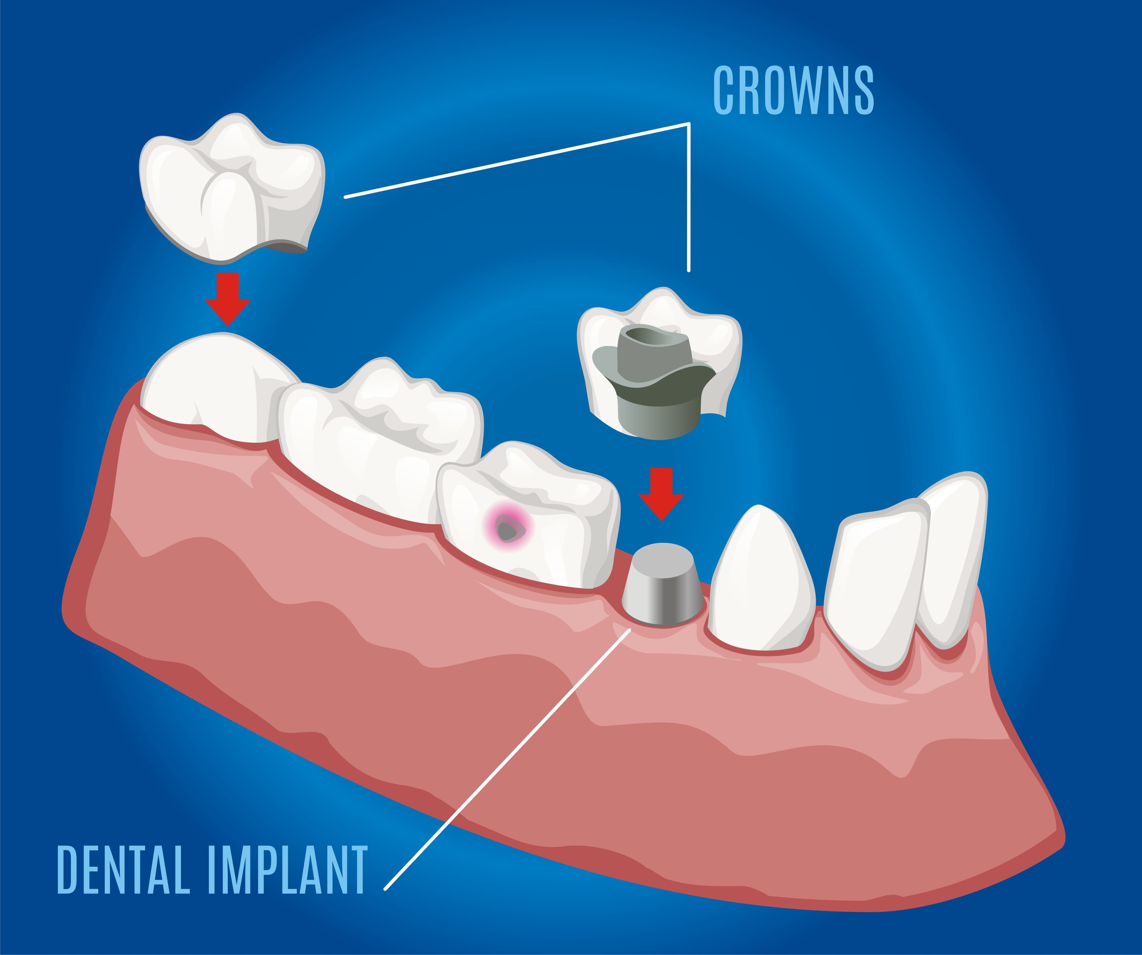
What Is a Dental Bone Graft?
A bone graft is the placement of bone material — from one of several sources — into the deficient area to stimulate new bone formation. It's not a replacement structure that stays forever. It's a scaffold. The body's own bone-forming cells (osteoblasts) use the graft material as a framework, remodel it, and replace it with the patient's own new bone over 3 to 6 months.
The graft itself dissolves and is absorbed as new bone fills in. What remains after healing is the patient's own bone — denser, more voluminous, and suitable for implant placement.
Types of Bone Graft Material
Several sources of bone material are used in dental grafting
Autograft (your own bone). The gold standard in terms of biological outcomes — the body accepts its own bone without risk of rejection or immune response, and new bone formation is predictably robust. The downside is a second surgical site to harvest bone from. Common donor sites in the mouth include the chin, the lower molar area, or the upper wisdom tooth region. For larger grafts, bone may occasionally be taken from outside the mouth (hip or tibia), but this is rarely needed in routine dental bone grafting.
Allograft (donor human bone). Bone from a processed human tissue bank — sterilised, freeze-dried, and rendered inert before use. It serves as a scaffold for the patient's own bone to grow into. No second surgical site is needed. It's widely used and clinically effective.
Xenograft (animal bone — typically bovine). Processed bovine bone that has had all organic components removed, leaving the mineral scaffold. Bio-Oss is the most widely used commercial xenograft product. It integrates well, is gradually replaced by patient bone, and has decades of clinical evidence supporting its use.
Alloplast (synthetic bone substitutes). Artificially manufactured graft materials — including beta-tricalcium phosphate, hydroxyapatite, and bioactive glass. These dissolve and are replaced by bone over time. Useful when patient- or donor-sourced materials aren't appropriate.
In most routine implant-site bone grafting, allograft or xenograft materials are used — avoiding the second surgical site while providing an effective scaffold for bone regeneration. At Renew Dental Clinic, Dr. Suchi Singh selects the appropriate material based on the size of the defect, the patient's healing profile, and the planned implant timeline.
Common Situations Where a Bone Graft Is Needed
Socket preservation (immediate post-extraction grafting). When a tooth is extracted, packing graft material into the fresh socket before it's stitched significantly reduces the bone resorption that would otherwise occur. This isn't always necessary — but for patients planning an implant at that site, socket preservation at the time of extraction preserves bone volume and avoids the need for a larger graft later.
Ridge augmentation. For patients who had a tooth extracted years ago and have experienced significant bone resorption — the ridge is now too narrow or too short for an implant of appropriate dimensions. Graft material is placed against the outer surface of the bone (block grafting) or within an extracted defect, and a membrane is placed over it to protect the graft as it integrates.
Sinus lift (maxillary sinus augmentation). The upper back teeth sit directly below the maxillary sinuses — the air-filled cavities in the cheekbones. When upper back teeth are lost, the sinus floor can "drop" downward as bone resorbs, leaving insufficient vertical bone height for an implant without entering the sinus. A sinus lift places bone graft material between the sinus lining and the bone floor, raising the sinus floor and creating space for an implant above it. It's one of the most predictable bone grafting procedures in dentistry.
Grafting around existing implants (treatment of peri-implantitis). When an implant develops gum disease (peri-implantitis) and bone has been lost around it, grafting can be used to regenerate that bone and stabilise the implant.
The Bone Graft Procedure — What Actually Happens
Before surgery. A detailed 3D CBCT scan is taken to assess the exact dimensions of bone available, the position of anatomical structures (nerve canals, sinus floor), and to plan the graft precisely. This imaging is what separates planned, safe surgery from guesswork.
Anaesthesia. Local anaesthesia is used for all minor oral surgical grafting procedures. The area is completely numbed before any incision. You feel pressure and movement during the procedure, not pain.
The surgical procedure. An incision is made in the gum to expose the bone. The graft material is placed, shaped to fill the defect, and secured. In most cases, a collagen membrane is placed over the graft to protect it from being disrupted by the overlying gum tissue before new bone has formed (guided bone regeneration). The gum is then sutured closed.
Healing time. The graft needs 3 to 6 months to integrate before implant placement. During this period, the graft is left undisturbed while the body's bone-forming cells gradually replace it with new bone. For socket preservation at the time of extraction, implant placement is typically planned for 4 to 6 months later. For larger ridge augmentation, 6 months may be needed.
What Recovery Looks Like
Bone grafting involves more recovery than a routine extraction but is typically managed well at home.
First 48 hours: Swelling and bruising in the area are expected and normal. Ice packs to the cheek reduce both. Mild to moderate soreness is managed with prescribed pain relief. Soft diet strictly.
Days 3 to 7: Swelling begins to reduce. Soreness improves. Avoid any pressure on the graft site. No poking, probing, or disturbing the stitches. Gentle salt water rinses after meals from day 2.
Weeks 2 to 4: Dissolvable sutures resolve. The graft site should feel comfortable. Soft or normal diet as tolerated. No strenuous exercise for the first two weeks.
Months 1 to 6: This is the bone formation period. The area may look and feel completely normal, but internally new bone is forming. A follow-up CBCT scan at the appropriate time confirms adequate bone formation before implant placement.
What to avoid during the healing period:
- Smoking — nicotine dramatically impairs bone formation and graft integration
- Alcohol — interferes with healing and the body's response to surgery
- Probing or disturbing the graft site
- Hard foods on the surgical side
What If I Had a Graft and the Implant Still Didn't Work?
Bone grafts, like any biological process, can occasionally fail to produce adequate bone. This is most common in heavy smokers, patients with poorly controlled diabetes, patients on bisphosphonate medications (for osteoporosis), and those who don't follow aftercare properly.
If a graft doesn't produce sufficient bone, re-grafting is an option — though the timeline extends considerably. Patients considering implants who smoke should ideally stop before grafting to give the procedure the best chance of success.
Frequently Asked Questions
1. Does getting a bone graft mean I'm definitely getting an implant?
Bone grafting is almost always done in preparation for an implant. Occasionally it's done to preserve bone after extraction even when the implant decision isn't yet made — socket preservation at the time of extraction is low-effort and high-value if an implant is a future possibility.
2. Will I feel any pain during the graft?
Not during the procedure, which is performed under local anaesthesia. Post-operative soreness is expected for several days and is managed with medication.
3. How do I know if I need a bone graft?
Only a clinical assessment with appropriate imaging (OPG X-ray and ideally a 3D CBCT scan) can determine whether you have sufficient bone for implant placement without grafting. Many patients assume they need a graft and don't; some assume they don't and do.
4. Can I eat normally after a bone graft?
Soft diet for the first 1 to 2 weeks. Return to normal eating as the area heals, avoiding hard food on the graft side.
Considering an Implant? Start with an Assessment at Renew Dental, Noida
Whether you've been told you need a bone graft or simply want to know whether you qualify for an implant — Renew Dental Clinic in Sector 47, Noida provides the imaging assessment and clinical evaluation needed to give you a clear, honest answer.
To book a consultation, call (0120) 498-8333.
Monday–Saturday 10:30 AM – 8:00 PM | Sunday 11:00 AM – 2:30 PM.
